Tomosynthesis
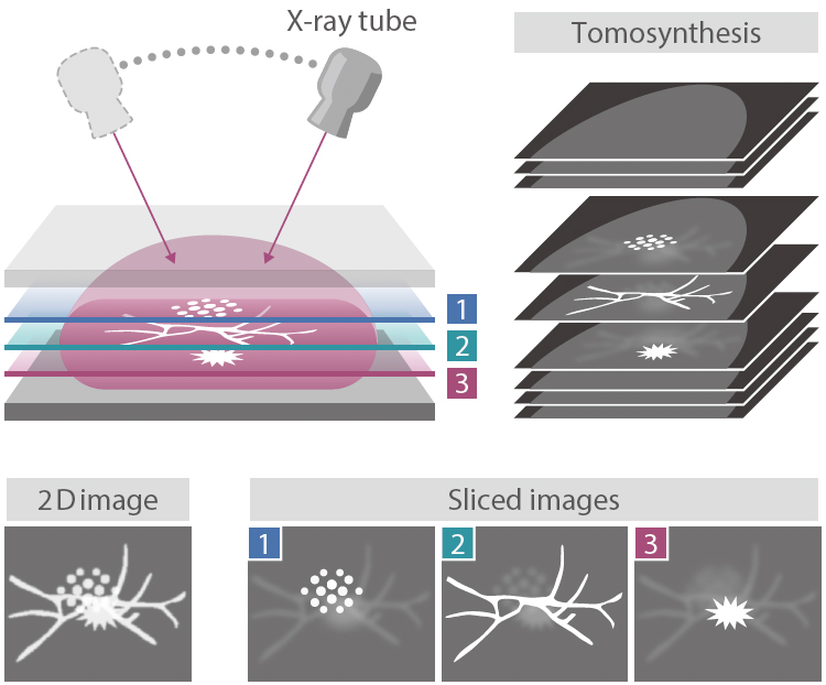
making it possible to observe the internal structure of the breast
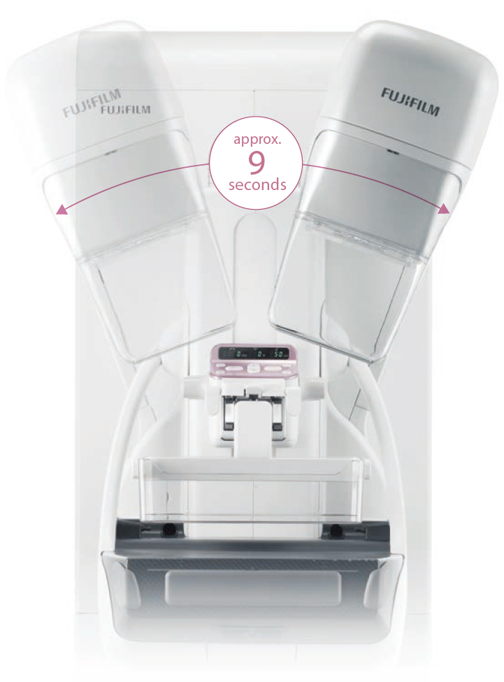
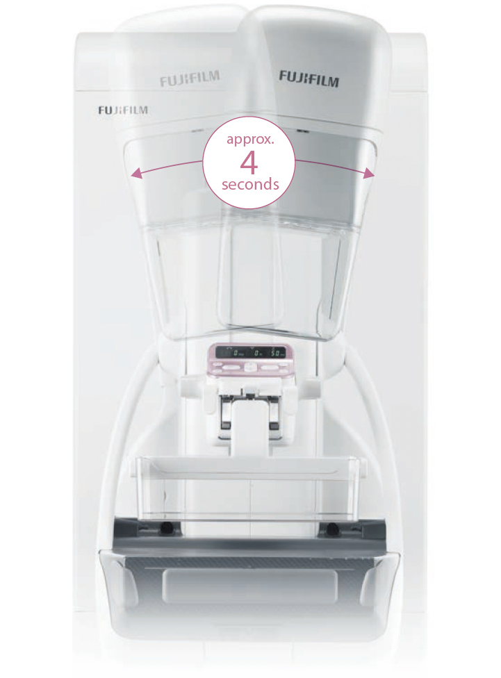
In breast tomosynthesis, the X-ray tube moves through an arc while acquiring a series of low-dose X-ray images.
The images taken from different angles are reconstructed into a range of Tomosynthesis slices where the structure of interest is always in focus.
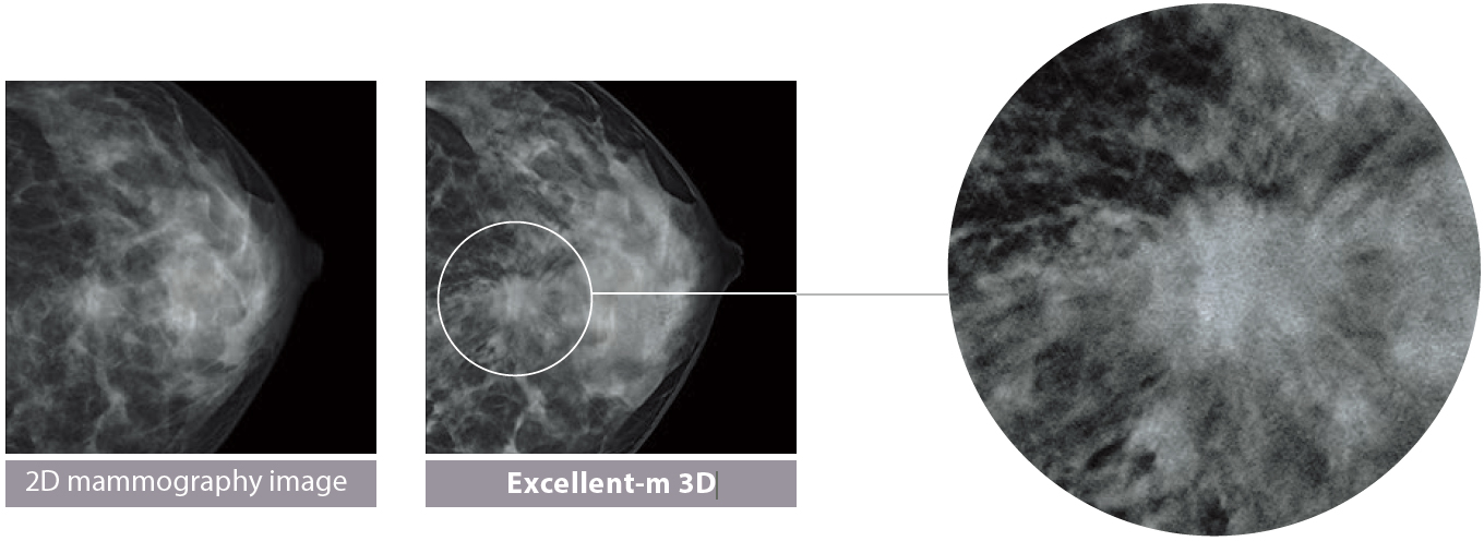
The reconstructed tomographic images make it easier to identify lesions which might be difficult to visualize in routine mammography because of the presence of overlapping breast structures.
The Tomosynthesis function on AMULET Innovality is suitable for a wide range of uses, offering two modes to cater for various clinical scenarios. Standard (ST) mode combines rapid exposure timing and efficient workflow with a low X-ray dose while High Resolution (HR) mode makes it possible to produce images with an even higher level of detail, allowing the region of interest to be brought into clearer focus.
S-View(synthesized 2D image)function is available
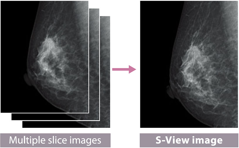
Tomosynthesis by AMULET Innovality automatically produces not only tomograms obtained at 1 mm intervals but also a two- dimensional S-View image combining multiple slice images.
With the S-View image showing the overall view added to tomograms offering the views in detail, comprehensive image reading is possible.

1. Reducing graininess of image in low-dose tomography
The image patterns are recognized to selectively suppress the patterns that do not exist in human body architectures as noise, to reduce distractive noises in the event of low-dose tomography.
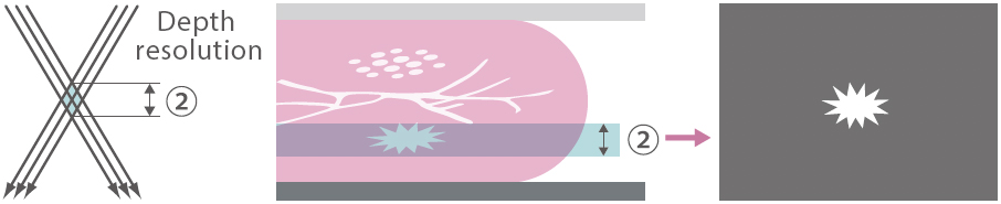
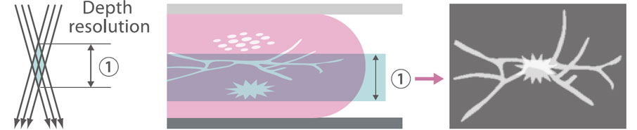
2. Suppressing interference of human body architectures at different depths (as illustrated on the right)
In the process of reconstructing the 3D breast architecture from multiple 2D images, calcification, mass, spicula, mammary gland and other signals that emerge from different depths in the breast architecture are selected off to reproduce the breast architecture at the focus depth with greater fidelity.
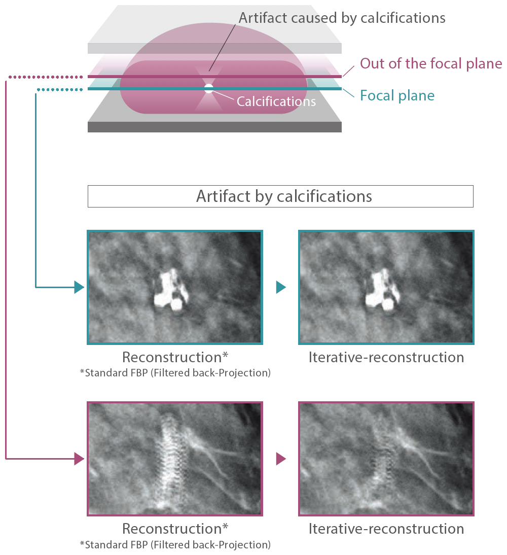
3. Restoring the fine-structure
Our super-resolution technology is introduced to restore the fine-structure of calcification and other phenomena, the visibility of which is impaired by the movement of the X-ray tube, to facilitate interpretation of tomosynthesis images.
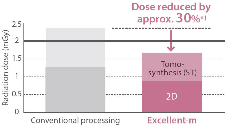
Offers significantly lower doses than the conventional method
With combination of 2D and Tomosynthesis Dose of 2 or less mGy is available*2
*1 Equivalent to an image of 40 mm PMMA compared with previous images (Breast thickness of 45 mm, 50% mammary gland, 50% fat)
*2 IAEA guidance level: 3 mGy, guidelines of the Japan Association of Radiological Technicians: 2 mGy
*In-house comparison
Two modes suitable for a range of clinical purposes
HR (High Resolution) mode
Acquisition angle: ±20° Pixel size: 100/50 mWith a larger acquisition angle the depth resolution is improved. This allows the region of interest to be defined more clearly and brought into clearer focus.
Additional imaging for complete checkup, grasping morphology, etc.
ST (Standard) mode
Acquisition angle: ±7.5°
Pixel size: 100/150 m The smaller angular range and fast image acquisition allow Tomosynthesis scans to be quickly performed with a relatively low X-ray dose.
Check-up, screening, follow-up, etc.
Static face guard for Tomosynthesis imaging (Face Guard T Comfort)
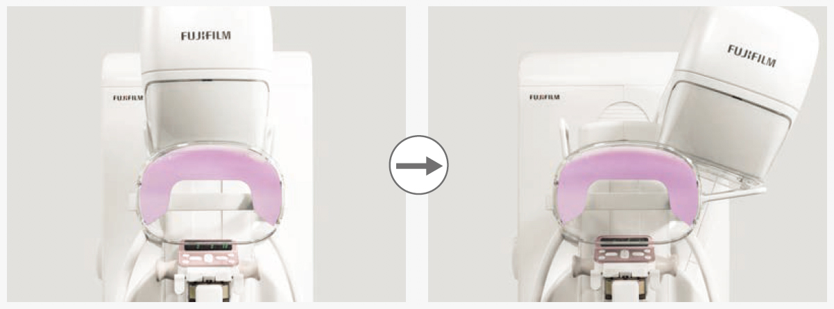
Fixing the face guard to the device instead of the tube part eliminates movement of the face guard during Tomosynthesis imaging. It will not be reflected at any angle of the ST mode (15 degrees) or HR mode (40 degrees). It can also be used as-is for normal mammography imaging.
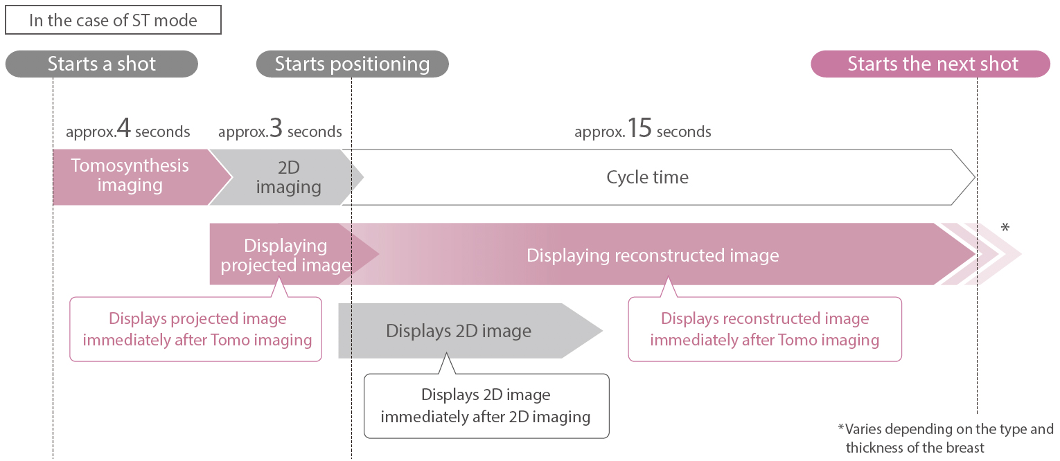
Shortens the imaging cycle with a fast display and reconstruction
After a shot, the next shot in either 2D or 3D can be started with a cycle time of approx. 15 seconds.